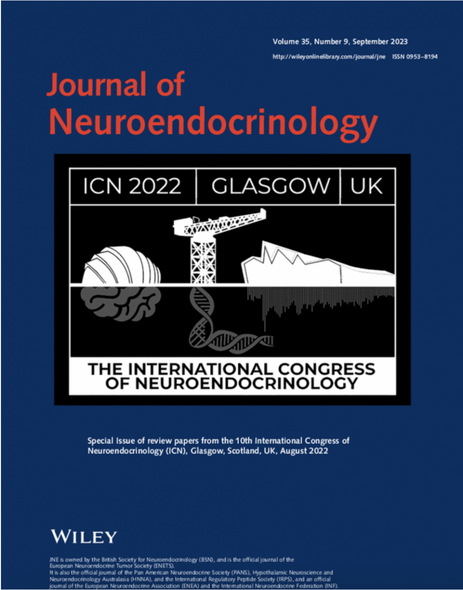
Journal of Neuroendocrinology Special Issue – International Congress of Neuroendocrinology 2022
 We are delighted to announce the publication of a Special Issue of articles from the International Congress of Neuroendocrinology 2022.
The special issue comprises an editorial by Editor-in-Chief Julian Mercer, 11 reviews and 1 original research article. All articles are either open access or available for free for 2 months.
The ICN is held every four years and is the biggest international meeting for neuroendocrinologists across the globe. The 2022 Congress was held in Glasgow, UK.
Browse the Special Issue: onlinelibrary.wiley.com/toc/13652826/2023/35/9
Contents
EDITORIAL
ICN 2022: Meeting up again and celebrating neuroendocrinology with the international community - Julian Mercer
REVIEW ARTICLES
Targeting carnitine palmitoyltransferase 1 isoforms in the hypothalamus: A promising strategy to regulate energy balance – OPEN ACCESS
Rosalía Rodríguez-Rodríguez, Anna Fosch, Jesús Garcia-Chica, Sebastián Zagmutt, Nuria Casals
Up- and downstream effectors of brain CPT1 isoforms for bodyweight control. CPT1A and CPT1C functions are tightly regulated by AMP-activated protein kinase (AMPK)/malonyl-CoA axis. In the hypothalamus, malonyl-CoA levels are attenuated by fasting or ghrelin, and are raised in response to refeeding, glucose, or leptin. Changes in malonyl-CoA levels are sensed by CPT1C, which, in turn, regulates the activity in neurons of other effectors such as ABHD6, an endocannabinoid (eCB) and SAC1, a PI4P phosphatase, to modulate peripheral metabolism and metabolic flexibility. Increased malonyl-CoA levels inhibit CPT1A catalytic activity to attenuate fatty acid oxidation (FAO), leading to long-chain fatty acid (LCFA) accumulation in the hypothalamus, resulting in a satiety signal that controls liver glucose production. MCD, malonyl-CoA descarboxylase.
Microbes, oxytocin and stress: Converging players regulating eating behavior – OPEN ACCESS
Cristina Cuesta-Marti, Friederike Uhlig, Begoña Muguerza, Niall Hyland, Gerard Clarke, Harriët Schellekens
The effects of central oxytocin on eating behavior, social behavior and stress and potential interaction of the gut microbiota. Oxytocin (Oxt) is a neuropeptide that is produced in magnocellular neurons (MCN) located in the paraventricular nucleus (PVN) and supraoptic nucleus (SON) of the hypothalamus (HYP) as well as in parvocellular neurons (PCN) located in the PVN (blue box). The arcuate nucleus (ARC, blue region) express OXTR and project to neurons in the PVN (orange arrow). Oxytocinergic neurons from the PVN (pink lines) project to many other brain regions (pink arrows) that are involved in the control of eating behavior including the nucleus tractus solitarius (NTS), the ventral tegmental area (VTA), the hippocampus (HPC), the amygdala (AMY), the nucleus accumbens (NAcc), and the prefrontal cortex (PFC). Brain regions with Oxt neurocircuitry have been shown to be involved in the control of stress (green circle), social behaviors (pink/red circle) and food intake behavior (yellow circle). The gut microbiota (purple circle) has been linked (blue arrows) with stress, eating behavior and Oxt independently, but also with Oxt particularly in social behavior context (the three effects are represented within a pyramid). The vagus nerve (in orange) connects the gut and the brain via gut-brain axis and is one mechanism by which gut microbiota is suggested to regulate appetite, eating and social behavior and stress.
Eat, seek, rest? An orexin/hypocretin perspective – OPEN ACCESS
Daria Peleg-Raibstein, Paulius Viskaitis, Denis Burdakov
Hypocretin/orexin neurons as a nutrient-gated switch between appetitive and consummatory behaviour. Some nutrients (e.g., non-essential amino acids, nAAs) activate HONs thus inducing a suppression of eating and reingnition of exploration (appetitive behaviour). Other nutrients (glucose) suppress HONs, thus facilitating consummatory behaviours as well as “rest and digest” physiology.
Adverse effects of gestational ω‐3 and ω‐6 polyunsaturated fatty acid imbalance on the programming of fetal brain development – OPEN ACCESS
Valentina Cinquina, Erik Keimpema, Daniela D. Pollak, Tibor Harkany
Cellular enrichment of the cerebral cortex during fetal brain development, including the timeline of neuronal and glial diversification, and the formation of neuronal networks. Major bioenergetic sources and potential modifications inflicted upon cellular identity were also marked.
Serotonergic regulation of appetite and sodium appetite
Yurim Shin, Seungjik Kim, Jong-Woo Sohn
This review article summarizes the role of central serotonin system in the regulation of appetite and sodium appetite.
Stress and the risk of Alzheimer dementia: Can deconstructed engrams be rebuilt?
Freddy Jeanneteau
Timelapse imaging of fluorescent tracers during AD progression reveals a spine attrition focus at amyloid plaques surrounded by a halo of spinogenesis consistent with the abnormally high proportions of silent neurons proximal to plaques and hyperactive neurons in its surroundings.
Pantelis Antonoudiou, Bradly Stone, Phillip L. W. Colmers, Aidan Evans-Strong, Najah Walton, Jamie Maguire
Stress and fear alter network states in emotional processing hubs. Theta oscillations (4-8 Hz) increase within and between the BLA, PFC, and HPC in response to stress and fear.Gamma oscillations (40 – 120 Hz) increase in the BLA and decrease in the PFC in association with stress and fear behaviors. Understanding the dynamics in network states related to stress and fear can further our understanding of how communication within and between brain regions drives specific behavioral states.
Rae Silver, Yifan Yao, Ranjan K. Roy, Javier E. Stern
The pituitary portal system was first described, almost 100 years ago, in human tissue using a hematoxylin and eosin stain (left). A second portal system, between the brain clock in the suprachiasmatic nucleus and the organum vasculum lamina terminalis was discovered using iDisco-cleared mouse brain stained with arginine vasopressin to identify SCN and collagen to label blood vessels (right). Today's tools permit not only a finer depiction of these blood vessels but also more certainty about their anatomical features.
ORIGINAL RESEARCH
Zahra S. Thirouin, Claire Gizowski, Anzala Murtaz, Charles W. Bourque
The circadian pattern of action potential discharge by vasopressin neurons of the suprachiasmatic nucleus shows a greater amplitude in males compared to female rats.
REVIEW ARTICLES
Social zebrafish: Danio rerio as an emerging model in social neuroendocrinology
Kyriacos Kareklas, Magda C. Teles, Ana Rita Nunes, Rui F. Oliveira
The role of neuroendocrine systems in the evolution of social phenotypes remains underexplored. We review evidence supporting the utility of zebrafish, Danio rerio, in providing impactful progress in this field. We discuss three key social phenotypes: social dominance, social affiliation and social cognition. Neuroplasticity and functional connectivity between brain areas are leading drivers of behavioural changes, which are chiefly modulated by the nonapeptide system and interactions with reward-pathway signalling, sex hormones and stress physiology.
Takumi Oti, Hirotaka Sakamoto
In this report, we review the neural regulatory mechanisms of the sexually dimorphic functions in the central nervous system and these neuropeptides on male sexual behaviour. Furthermore, we discuss the finding of a recently identified, localised “volume transmission” role of oxytocin in the spinal cord.
KNDy neurones and GnRH/LH pulse generation: Current understanding and future aspects
Uncovering the central mechanism underlying mammalian reproduction is warranted to develop new therapeutic approaches for reproductive disorders in humans and domestic animals. This review focused on the role of arcuate kisspeptin/neurokinin B (NKB)/dynorphin A (Dyn)-co-expressing neurones (also known as KNDy neurones) as an intrinsic gonadotropin-releasing hormone (GnRH) pulse generator, which plays a fundamental role in mammalian reproduction. We also discuss the current understanding of the mechanism inhibiting pulsatile GnRH/gonadotropin release during malnutrition and provide the aspects on the seeds of reproductive drugs for hypothalamic reproductive disorders in humans and livestock.

